


The human heart is an electrical marvel, and capturing its rhythm requires more than just a high-tech machine; it requires meticulous documentation. Whether you are a medical student, a biomedical engineer, or a clinical researcher, maintaining a detailed Ecg components lab notebook is the bridge between seeing a line on a screen and truly understanding cardiac health. In this guide, we will dive deep into the nuances of electrocardiography, focusing on how to systematically record and analyze every wave, segment, and interval to ensure your lab records are professional and clinically accurate.
- Why You Need a Dedicated Ecg Components Lab Notebook
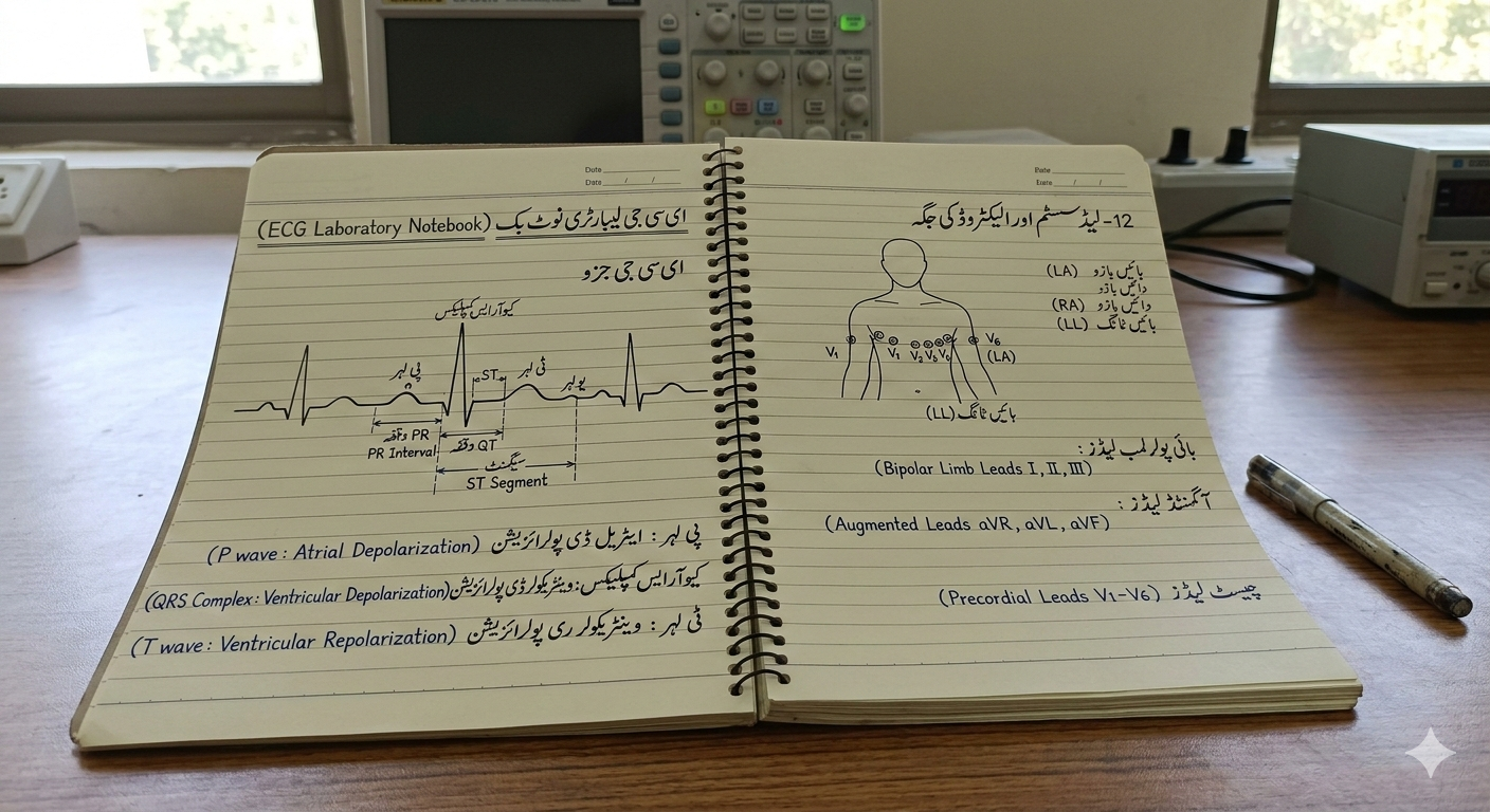
- The Anatomy of the Wave: Core Entries for Your Notebook
- The P Wave: Atrial Depolarization
- The QRS Complex: Ventricular Power
- The T Wave: Recovery and Repolarization
- Measuring Intervals: The Math of the Ecg Components Lab Notebook
- Organizing Your Ecg Components Lab Notebook for Success
- Advanced Observations: Segments and Baselines
- Digital vs. Paper: Choosing Your Lab Notebook Style
- Common Pitfalls in ECG Documentation
- Conclusion: Turning Data into Insight
- Frequently Asked Questions (FAQs)
- 1. What is the most important component to record in an Ecg components lab notebook?
- 2. How do I measure intervals if the baseline is wandering?
- 3. Should I include “R-R intervals” in my lab notebook?
- 4. What are the “small boxes” and “large boxes” on ECG paper?
- 5. Can I use a regular notebook, or do I need a specialized “Ecg components lab notebook”?
Why You Need a Dedicated Ecg Components Lab Notebook
In a world dominated by digital health records, the art of the physical or digital Ecg components lab notebook remains a vital skill. Documentation isn’t just about keeping a history; it’s about developing the “clinical eye” required to spot minute abnormalities that automated software might overlook. By manually logging components, you force your brain to process the morphology of each wave, which builds long-term diagnostic intuition.
A well-organized notebook serves as a personal reference library. When you encounter a rare arrhythmia or a subtle ST-segment elevation, having a structured history of previous logs allows you to compare data points across different subjects or timeframes. It transforms a chaotic stack of thermal paper strips into a cohesive narrative of cardiac physiology.
The Anatomy of the Wave: Core Entries for Your Notebook
Every entry in your Ecg components lab notebook should begin with the fundamental waveforms. To document these correctly, you must understand what each deflection represents in the cardiac cycle. Let’s break down the essential “big three” that form the backbone of your lab observations.
The P Wave: Atrial Depolarization
The P wave is the first deflection you will record. It represents the electrical impulse as it travels from the SA node through the atria. In your Ecg components lab notebook, you should note its amplitude (usually less than 2.5 mm) and its duration (usually less than 0.11 seconds).
If the P wave is notched or peaked, these are critical observations. A notched P wave often suggests left atrial enlargement, while a tall, peaked P wave may point toward right atrial issues. Recording these shapes precisely is what separates a basic log from a professional-grade lab notebook.
The QRS Complex: Ventricular Power
The QRS complex is the most “famous” part of the ECG, reflecting the rapid depolarization of the right and left ventricles. Because the ventricles have a much larger muscle mass than the atria, the QRS is significantly larger than the P wave.
In your documentation, measure the QRS duration carefully. A normal complex is narrow (0.06 to 0.10 seconds). If you find a wide QRS in your Ecg components lab notebook, it indicates that the impulse is taking a “scenic route” through the ventricles, potentially signaling a bundle branch block or a ventricular origin of the beat.


The T Wave: Recovery and Repolarization
The T wave represents ventricular repolarization. While it might look like a simple bump, it is perhaps the most sensitive component to metabolic changes. In your lab notes, pay attention to T-wave inversion or “tenting.” Tall, peaked T waves are often the first sign of hyperkalemia (high potassium), making this a high-priority data point for your records.
Measuring Intervals: The Math of the Ecg Components Lab Notebook
Beyond the waves themselves, the timing between the waves tells the real story. To fill your Ecg components lab notebook with high-quality data, you need to master the use of calipers or the “box counting” method.
The PR Interval: The Delay is Key
The PR interval measures the time from the start of atrial depolarization to the start of ventricular depolarization. It’s essentially a measurement of how long the AV node holds the signal.
-
Normal range: 0.12 to 0.20 seconds.
-
Lab Note: If this interval is consistently long, you are likely looking at a first-degree heart block.
The QT Interval: A Critical Safety Marker
The QT interval encompasses both ventricular depolarization and repolarization. It is highly dependent on heart rate, so your Ecg components lab notebook should always include the “corrected” QT (QTc).
-
Why it matters: A prolonged QT interval is a red flag for potential lethal arrhythmias like Torsades de Pointes.
-
Documentation Tip: Always record the heart rate alongside the QT to ensure your calculations are contextually accurate.
Organizing Your Ecg Components Lab Notebook for Success
A messy notebook is a useless notebook. To maintain a professional standard, you should follow a consistent template for every entry. This ensures that you don’t skip critical steps during high-pressure lab sessions.
1. Subject Metadata
Before looking at the waves, record the basics:
-
Subject ID/Age/Sex.
-
Date and Time of the recording.
-
Current medications (especially beta-blockers or anti-arrhythmics).
-
Physical state (e.g., resting, post-exercise, or stressed).
2. The Lead Analysis
An ECG is a 3D view of a 3D heart recorded on 2D paper. Your Ecg components lab notebook should specify which lead you are analyzing. Lead II is the standard for rhythm strips because it follows the heart’s natural electrical axis, but the precordial leads (V1-V6) are essential for localized muscle analysis.
3. Quantitative Data Table
Create a small grid in your notebook for every entry: | Component | Duration (s) | Amplitude (mV) | Morphology Notes | | :— | :— | :— | :— | | P Wave | 0.08 | 0.1 | Smooth, upright | | PR Interval | 0.16 | N/A | Constant | | QRS Complex | 0.09 | 1.2 | Narrow, R-wave progression normal | | ST Segment | N/A | 0 | Isoelectric |
Advanced Observations: Segments and Baselines
While intervals include the waves, segments are the flat lines between them. The most important segment for your Ecg components lab notebook is the ST segment.
The ST segment should ideally sit on the isoelectric line (the baseline). If you notice the ST segment is elevated or depressed, this is a “stop everything” moment in a clinical setting, as it often indicates myocardial ischemia or injury.
In a lab notebook setting, always describe the ST segment in relation to the TP segment (the interval between the end of the T wave and the next P wave). Using the TP segment as your baseline reference is the gold standard for accuracy in electrocardiography.
Digital vs. Paper: Choosing Your Lab Notebook Style
There is an ongoing debate about whether a physical Ecg components lab notebook or a digital tablet-based system is better.
The Case for Paper:
-
Tactile Learning: Drawing the waveforms helps with retention.
-
No Battery Issues: Your notebook won’t die in the middle of a 12-hour shift.
-
Easy Annotation: You can quickly circle a PVC or an U-wave without navigating menus.
The Case for Digital:
-
Searchability: You can find “Patient 402” in seconds.
-
Image Integration: You can paste a digital scan of the ECG strip directly next to your analysis.
-
Collaboration: Easily share your findings with a supervisor or colleague for a second opinion.
Whichever medium you choose, the consistency of your Ecg components lab notebook is what determines its value.
Common Pitfalls in ECG Documentation
Even experienced researchers can make mistakes when filling out their Ecg components lab notebook. Awareness of these common errors will help you maintain the integrity of your data.
-
Ignoring Artifacts: Muscle tremors, loose electrodes, or 60-cycle interference can create “waves” that aren’t real. Always note “artifact present” in your logs to avoid misdiagnosing a wandering baseline as a cardiac event.
-
Miscalculating Rate: Remember the 300-150-100-75-60-50 rule for regular rhythms, but use the 6-second strip method for irregular rhythms. Mixing these up in your notebook can lead to incorrect heart rate data.
-
Neglecting the U Wave: Sometimes a small wave follows the T wave. While often normal, it can indicate low potassium. Don’t leave it out of your Ecg components lab notebook just because it’s small!
Conclusion: Turning Data into Insight
The Ecg components lab notebook is more than just a collection of numbers and scribbles; it is a vital tool for mastering the language of the heart. By systematically documenting the P, QRS, and T waves, along with their respective intervals and segments, you develop a disciplined approach to cardiac diagnostics.
Whether you are tracking the progress of a clinical trial or studying for your medical boards, the rigor you apply to your notebook today will pay dividends in your clinical accuracy tomorrow. Start your notebook with a clear template, stay consistent with your measurements, and always look beyond the primary waves to find the hidden stories in the segments and intervals.
Next Step: Why not try creating your first template page? Grab a ruler, set up your columns for waves and intervals, and practice logging a “Normal Sinus Rhythm” to set your baseline!
Frequently Asked Questions (FAQs)
1. What is the most important component to record in an Ecg components lab notebook?
While all components are vital, the QRS complex is often considered the most critical because it reflects ventricular activity. However, for a complete clinical picture, the ST segment is equally important as it is the primary indicator of acute cardiac injury or ischemia.
2. How do I measure intervals if the baseline is wandering?
A wandering baseline is common in a lab setting. To measure intervals accurately in your Ecg components lab notebook, use the TP segment (the space between the T wave and the next P wave) as your reference point. If the wander is too severe, check the electrode contact and restart the recording.
3. Should I include “R-R intervals” in my lab notebook?
Absolutely. Measuring the distance between consecutive R waves is essential for determining rhythm regularity. If the R-R interval varies significantly, you must document this as an “irregularly irregular” or “regularly irregular” rhythm in your logs.
4. What are the “small boxes” and “large boxes” on ECG paper?
On standard ECG paper, one small box represents 0.04 seconds and 0.1 mV. One large box (consisting of 5 small boxes) represents 0.20 seconds. Mastering these units is fundamental to making accurate entries in your Ecg components lab notebook.
5. Can I use a regular notebook, or do I need a specialized “Ecg components lab notebook”?
While a specialized notebook with grid-printed pages is helpful for pasting strips, a regular notebook works fine as long as you maintain a consistent, structured template. The value lies in your methodology and the accuracy of your measurements, not the brand of the notebook.